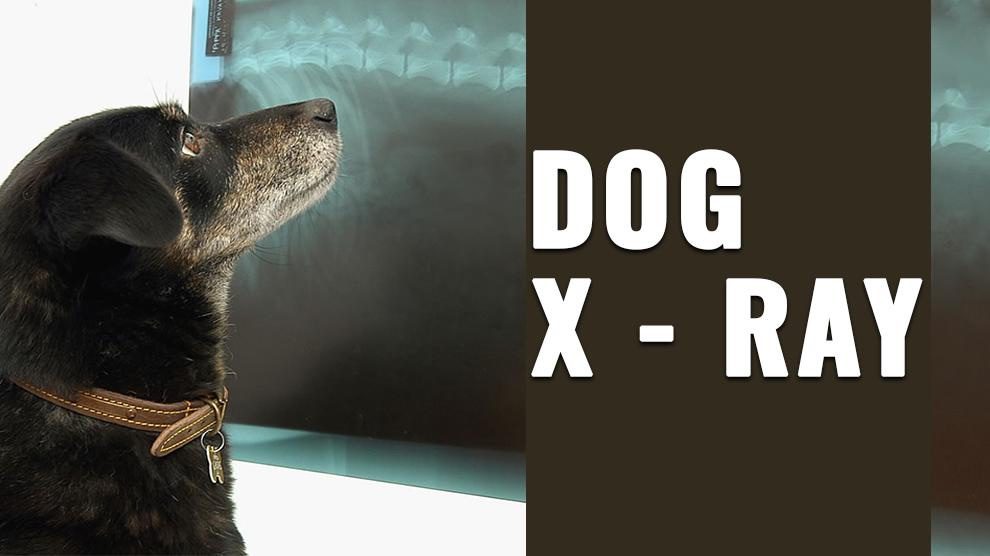
- How Much Is An X-Ray For A Dog?
- X-Rays For Pregnant Dogs
- Dog Knee X-Ray
- X-Ray Of Dogs Stomach
- Dog Dental X-Ray
- How Do Dogs Get X-Rays?
- X-Ray Of Dog With Lung Cancer
- X-Ray Dog Abdomen
- X-Ray Dog Hip Dysplasia
- X-Ray Of Dog Tail
- Dog Leg X-Ray
- X-Ray Vs Ultrasound For Dogs
- Dog X-Ray Machine
- Difference Between Ultrasound, X-Ray, CT Scan, MRI
- Dog X-Ray FAQs

Dog Pregnancy Calculator And Timeline
Dogs are beloved companions and members of our family. They are also prone to various health issues just like humans. X-rays are an essential tool in diagnosing many of these health problems in dogs. They are frequently used to evaluate injuries, illnesses, and diseases that affect a dog’s bones, joints, lungs, and other internal organs.
While X-rays are generally considered safe for dogs, there are certain risks associated with the procedure, including radiation exposure.
These imaging tests use radiation to create detailed images of the internal structures of the body, helping veterinarians diagnose and treat a wide range of health problems. Dog X-rays are safe and painless and can be performed quickly, making them an essential tool in veterinary medicine.
Different body tissues absorb X-rays differently, resulting in varying levels of brightness on the X-ray image. Bones appear white, air and gas appear black and soft tissues appear shades of gray.
How Much Is An X-Ray For A Dog?
The cost of a dog X-ray can vary depending on several factors, including the type of X-ray, the location of the veterinary clinic, and the dog’s size. Generally, the cost of a dog X-ray ranges from $75 to $300, with an average of $150.
However, the cost can be much higher for more complex procedures, such as a full-body X-ray, or specialized imaging, such as CT scans or MRI. Pet insurance may cover a portion of the cost of a dog X-ray, depending on the plan’s coverage.
X-Rays For Pregnant Dogs
X-rays for pregnant dogs can be a valuable diagnostic tool when used properly. However, due to the potential risks associated with radiation exposure, it is important to use caution when using x-rays on pregnant dogs.
X-rays can be used during pregnancy to check the number and position of the puppies, monitor fetal growth, and identify any potential problems such as abnormalities or fetal distress.
However, the use of x-rays should be limited to situations where the information obtained is necessary for the health of the mother or puppies. Exposure to radiation during pregnancy can potentially harm the developing puppies, as well as the mother.
Therefore, it is important to limit the number of x-rays taken and to use appropriate safety measures, such as shielding the abdomen, to minimize radiation exposure.
In general, x-rays should only be used during pregnancy when absolutely necessary and under the guidance of a veterinarian.
Your veterinarian can help determine the best course of action based on your dog’s individual circumstances and the potential risks and benefits of using x-rays during pregnancy.
Dog Knee X-Ray
A dog knee X-ray, also known as a radiograph of the stifle joint, may be necessary if a dog is experiencing lameness or pain in their hind legs. The X-ray can help diagnose conditions such as cruciate ligament tears, patellar luxation, and osteoarthritis.
The cost of a dog knee X-ray can vary depending on the location, the size of the dog, and the veterinary clinic. Generally, the cost of an X-ray can range from $100 to $300.
However, additional fees may apply if sedation or anesthesia is required. Whether a dog knee X-ray is needed will depend on the specific symptoms and the veterinarian’s assessment.
A thorough physical exam and medical history will help determine if an X-ray is necessary. In some cases, the veterinarian may also recommend additional diagnostic tests such as blood work or joint fluid analysis.
A dog knee X-ray is important for proper diagnosis and treatment planning. It can help identify the underlying cause of the dog’s lameness or pain and guide the veterinarian in selecting the most appropriate treatment option, which may include medication, physical therapy, or surgery.
X-Ray Of Dogs Stomach
A stomach X-ray is used to evaluate a dog’s digestive system and identify any abnormalities, such as foreign objects or blockages.
This type of X-ray may be recommended if a dog has swallowed something that is not passing through the digestive system normally, or if the dog is experiencing symptoms of vomiting, abdominal pain, or diarrhea.
To perform a stomach X-ray, the dog is placed on its back on a table, and a lead apron or collar is placed over the dog’s chest to limit radiation exposure to the rest of the body.
The veterinarian or technician will then take several images of the dog’s abdomen and digestive system.
Dog Dental X-Ray
Dental X-rays are a valuable tool in veterinary dentistry and are commonly used to diagnose dental problems such as cavities, tooth root abscesses, and gum disease.
This type of X-ray can help veterinarians assess the health of the dog’s teeth and identify any underlying conditions that may not be visible during a routine dental exam.
To perform a dental X-ray, the dog is typically sedated to ensure that they remain still and to minimize any discomfort. The X-ray machine is positioned next to the dog’s mouth, and specialized dental X-ray film or digital sensors are used to capture the image.
How Do Dogs Get X-Rays?
The X-ray process for dogs is similar to that of humans. The dog is placed on a table or held still by a technician while the X-ray is taken. The X-ray machine emits a beam of radiation that passes through the dog’s body and onto a special film or digital sensor.
The image produced by the X-ray is then examined by a veterinarian or radiologist to diagnose any health conditions or injuries. To obtain the best quality X-ray image, the dog needs to remain still during the procedure.
This may require sedation for some dogs who are anxious or uncooperative. The dog’s hair may also need to be trimmed or shaved in the area being X-rayed to obtain a clearer image.
X-Ray Of Dog With Lung Cancer
Lung cancer is a serious health condition that can affect dogs. It is a common type of cancer in dogs, particularly in older dogs or those with a history of exposure to smoke or other pollutants.
An x-ray of a dog with lung cancer is a diagnostic test that can be used to evaluate the dog’s lungs and surrounding structures.
The x-ray images will show abnormalities in the lungs, such as the presence of tumors, fluid accumulation, and changes in the size and shape of the lungs.
The vet may also take additional images of the dog’s chest to evaluate the presence of cancerous cells in the lymph nodes or other organs.
To diagnose lung cancer with an x-ray, the dog is placed on the x-ray table and positioned in a way that allows the veterinarian to capture images of the chest. The images will show the size, shape, and location of any tumors or abnormalities in the lungs.
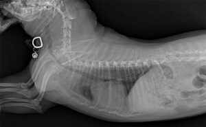
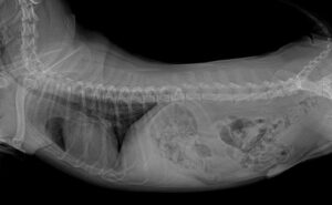
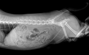
X-Ray Dog Abdomen
A dog x-ray abdomen is a diagnostic test that is used to evaluate the dog’s abdominal organs, such as the liver, spleen, kidneys, and intestines.
The x-ray can identify abnormalities in the size, shape, and location of these organs, which can be an indication of various health conditions.
The dog will need to lie still during the procedure, and the vet will take multiple images from different angles to get a comprehensive view of the organs.
X-Ray Dog Hip Dysplasia
Hip dysplasia is a common condition in dogs, particularly in large breeds, where the hip joint doesn’t form properly. It can cause pain, lameness, and arthritis, and can be diagnosed with an x-ray.
An x-ray of dog hip dysplasia is a diagnostic test that can be used to confirm the presence of this condition. The x-ray images will show abnormalities in the hip joint, such as the presence of bone spurs, joint space narrowing, and irregularities in the shape of the hip socket.
To diagnose hip dysplasia with an x-ray, the dog is placed on the x-ray table in a specific position that allows the veterinarian to evaluate the hips.
The dog’s legs are extended straight out behind it, and the x-ray machine is positioned above the hips. The resulting images will show the shape of the hip joint and any abnormalities, such as laxity or degenerative changes.
X-Ray Of Dog Tail
An x-ray of a dog’s tail is a diagnostic test that is used to evaluate the dog’s tailbone and surrounding structures. This type of x-ray is typically used to diagnose injuries or abnormalities in the tailbone, such as fractures, dislocations, or tumors.
This is particularly important in breeds with long tails, where injuries are more common. During the procedure, the dog is placed on the x-ray table in a position that allows the veterinarian to capture images of the entire tail.
The resulting images will show the shape and structure of the bones in the tail, as well as any abnormalities, such as fractures or tumors.
Dog Leg X-Ray
Leg X-rays are commonly used to diagnose orthopedic conditions such as fractures, joint problems, and ligament injuries.
This type of X-ray can help veterinarians identify the location and severity of the injury and determine the best course of treatment.
To perform a leg X-ray, the dog’s leg is typically positioned in a specific way to ensure that the X-ray captures the entire area of interest.
Depending on the dog’s size and the location of the injury, sedation may be necessary to ensure that the dog remains still during the procedure.
X-Ray Vs Ultrasound For Dogs
Both X-rays and ultrasound are commonly used diagnostic tools in veterinary medicine to evaluate the health of dogs. However, they have different applications and may be used for different purposes depending on the situation.
X-rays are best used to evaluate bones and joints, as well as the chest and abdominal cavities. They are particularly useful for detecting fractures, bone abnormalities, and changes in the size and shape of organs.
X-rays may also be used to identify foreign objects, such as rocks or toys, that a dog may have swallowed. Ultrasound, on the other hand, is a non-invasive imaging technique that uses high-frequency sound waves to create images of soft tissues and organs.
It is particularly useful for evaluating organs such as the heart, liver, kidneys, and bladder, as well as detecting fluid accumulation or masses. Ultrasound can also be used to guide biopsy procedures or evaluate pregnant dogs.
Top Rated Dog Trainers / Walkers / Breeders In Your Area
Dog X-Ray Machine
A dog x-ray machine is a specialized piece of equipment that is used to take x-ray images of a dog’s internal structures. This machine is similar to the x-ray machines used in human medicine, but it is designed to accommodate the size and shape of a dog.
The machine works by emitting a small amount of radiation that passes through the dog’s body and onto a photographic plate. The plate captures the image of the internal structures, which can be used to diagnose various health conditions.
The machine consists of a table on which the dog is placed, and an x-ray generator that produces the radiation used to create the images. The dog is positioned on the table in a way that allows the veterinarian to take the necessary images.
Once the dog is in position, the veterinarian will activate the x-ray generator, which produces a beam of radiation that passes through the dog’s body and onto a film or digital sensor.
The resulting image will show the bones, organs, and soft tissues inside the dog’s body.
Difference Between Ultrasound, X-Ray, CT Scan, MRI
Ultrasound, X-ray, CT scan, and MRI are all diagnostic imaging techniques, but they use different types of technology to create images and provide different types of information.
- Ultrasound uses high-frequency sound waves to create images of soft tissues and organs in real time. It is non-invasive, does not use radiation, and is often used to evaluate the heart, liver, kidneys, bladder, and other abdominal organs. It can also be used to guide biopsy procedures.
- X-rays use ionizing radiation to produce black-and-white images of bones and dense tissues such as the lungs and abdominal organs. X-rays are commonly used to detect fractures, foreign objects, and abnormalities in the chest and abdomen.
- CT (computed tomography) scan uses X-rays and advanced computer technology to create detailed 3D images of the body. CT scans are particularly useful for detecting internal injuries, fractures, and abnormalities in the chest, abdomen, and head. CT scans use a higher dose of radiation than X-rays.
- MRI (magnetic resonance imaging) uses a magnetic field and radio waves to create detailed images of soft tissues and organs. MRI provides high-resolution images of the brain, spine, joints, and soft tissues, and is particularly useful for detecting tumors, inflammation, and other soft tissue abnormalities. MRI does not use ionizing radiation but can be expensive and may require sedation or anesthesia.
Dog X-Ray FAQs
1. Do Dogs Need To Be Sedated For X-Ray?
It depends on the individual dog and the specific situation. In general, sedation is not always necessary for a dog to undergo an X-ray, but it may be recommended in certain circumstances.
For example, if a dog is very anxious or aggressive, sedation may be necessary to keep them calm and still during the procedure to ensure a clear and accurate image.
Additionally, if the X-ray requires a specific position, such as for orthopedic evaluations, sedation may be necessary to maintain the position.
However, sedation comes with risks, and veterinarians will carefully weigh the benefits versus the potential risks before recommending it.
Ultimately, the decision to sedate a dog for an X-ray will depend on the dog’s individual needs and behavior and the type of X-ray needed.
2. How Much Are Dog X-Rays On Leg?
The cost of dog x-rays on legs can vary depending on several factors, such as the location and size of the veterinary clinic, the specific type of x-ray needed, and any additional services that may be required.
In general, the cost of x-rays on a dog’s legs can range from $75 to $400 or more. If the x-ray is being done as part of a diagnostic workup for a specific health issue, additional fees may be charged for a consultation with the veterinarian or for any necessary follow-up tests or treatments.
Additionally, if sedation or anesthesia is required to keep the dog still during the x-ray, this may also increase the overall cost.
3. How Long Does A Dog X-Ray Take?
The length of time it takes to perform an X-ray on a dog can vary depending on a few factors, such as the size of the dog, the type of X-ray being performed, and the preparation needed for the X-ray.
In general, X-rays for dogs typically take between 10-30 minutes to complete. However, this can vary depending on the complexity of the X-ray and the cooperation of the dog during the procedure.
If the dog needs to be sedated or anesthetized for the X-ray, it can take longer as there will be additional time needed for the sedation to take effect and for the dog to recover afterward.
Additionally, if multiple views or angles are needed, it may take longer to complete the X-ray. It’s best to check with the veterinarian or the facility where the X-ray will be performed for a more accurate estimate of the time needed for the specific X-ray procedure.
4. How Much Is A Dog X-Ray Stomach?
The cost of a dog X-ray of the stomach can vary depending on the veterinary clinic, location, and other factors.
Generally, the cost can range from $100 to $300. It’s best to check with the specific clinic for an accurate estimate as prices may vary. Factors such as the region, size of the dog, and additional tests needed may also affect the cost.
5. Do X-Rays Show Stomach Problems?
X-rays can help identify some stomach problems in dogs, but they may not always provide a complete picture of the problem. When it comes to the stomach, X-rays can help detect issues such as foreign objects, obstructions, ulcers, and tumors.
However, X-rays may not be effective in identifying more subtle or non-specific stomach problems, such as inflammation, infections, or certain types of cancer.
In some cases, a series of X-rays may be necessary to track the movement of barium or other contrast materials through the digestive system, which can help identify issues with the stomach and intestines.
In addition to X-rays, other diagnostic tests such as blood work, ultrasound, endoscopy, or biopsy may also be necessary to fully evaluate a dog’s stomach problems.
A veterinarian may also evaluate a dog’s clinical signs and symptoms, and medical history, and perform a physical exam to determine the need for further testing.
6. Are X-Rays Bad For Dogs?
Dog X-rays are generally safe and do not cause any long-term harm to the dog. However, X-rays use ionizing radiation, which can be harmful in high doses. The amount of radiation exposure during a dog X-ray is relatively low and considered safe for dogs.
The risk of harm from radiation exposure is higher for pregnant dogs or puppies, so it is important to inform the veterinarian if the dog is pregnant or suspected of being pregnant.
To reduce radiation exposure, the dog may wear a lead apron or collar during the procedure. The veterinarian or technician may also take additional precautions to limit exposure to the radiation beam.
In general, the benefits of obtaining an accurate diagnosis from X-rays outweigh the small risk of radiation exposure.
7. Will An X-Ray Show A Tumor In A Dog?
An X-ray may show the presence of a tumor in a dog, but it depends on the type of tumor and its location. Tumors, which can be composed of different types of tissues, may appear as abnormal masses or growths on the X-ray image.
X-rays are useful in identifying tumors that involve bone, such as osteosarcomas, which can cause changes in the density or appearance of bone on the X-ray image.
However, X-rays are not always effective in detecting soft tissue tumors, such as lymphoma or mast cell tumors. Soft tissue tumors may not show up on an X-ray or may appear as a mass or swelling, which could be caused by other conditions as well.
In addition to X-rays, other diagnostic tests such as ultrasound, CT scan, MRI, or biopsy may be necessary to identify the presence and location of a tumor in a dog.
A veterinarian may also evaluate a dog’s clinical signs and symptoms, and medical history, and perform a physical exam to determine the need for further testing.
8. What Does Gas Look Like On An X-Ray?
On an X-ray, gas appears as black or dark areas. This is because X-rays are absorbed differently by different materials. Air and gas do not absorb X-rays as much as other tissues, such as bone or muscle, which makes them appear dark on the X-ray image.
When gas is present in the stomach or intestines, it can appear as black areas on the X-ray image. This can be helpful in diagnosing conditions such as gastrointestinal obstruction or foreign body ingestion, as the presence of gas can indicate the location and severity of the obstruction or foreign object.
However, it’s important to note that not all conditions or abnormalities will be visible on an X-ray. Other diagnostic tests such as ultrasound, endoscopy, or blood tests may also be necessary to fully evaluate a dog’s condition.